
X-Ray
HOW DOES X-RAY WORK?
X-rays are a type of radiation just like radio waves and light. The X-ray machine is positioned toward the part of the body being imaged. It then produces a small amount of radiation that passes through the body and captures an image on film or a special image recording plate.
WHAT SHOULD I EXPECT?
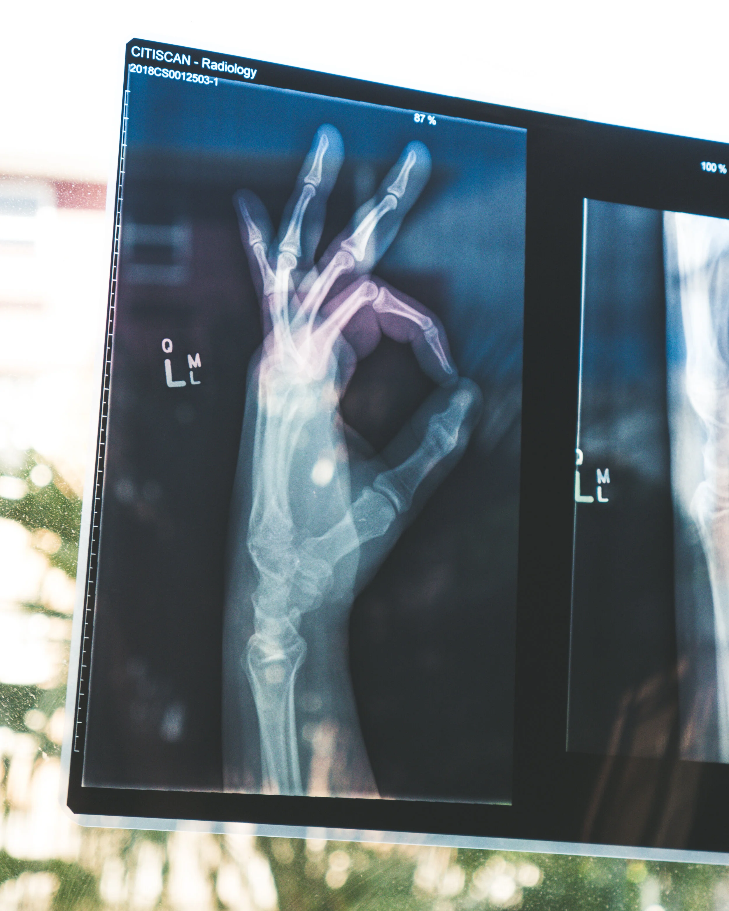
Radiography involves exposing a part of the body to a small dose of ionizing radiation to produce pictures of the inside of the body. X-rays, or radiation-like light or radio waves, pass through most objects, including the body. Once it is carefully aimed at the part of the body being examined, an X-ray machine produces a small burst of radiation that passes through the body, recording an image digitally. Different parts of the body absorb the X-rays in varying degrees. Dense bone absorbs much of the radiation while soft tissue, such as muscle, fat and organs, allow more of the X-rays to pass through them. As a result, bones appear white on the X-ray, soft tissue shows up in shades of gray and air appears black.
Generally, two or three X-rays will be taken depending on the body part that is being viewed. You will be asked to remain as still as possible during the very short exposure time. If necessary, you will be instructed to hold your breath in order to prevent motion from blurring the images. A patient may return to normal activities once the X-rays are complete.
HOW SHOULD I PREPARE?
There is no preparation for an X-ray exam, although patients may be asked to change into a gown to eliminate any interference from metal objects on clothing.
*Woman should always inform their physician and the X-ray technologist if there is any possibility of pregnancy.
OBTAINING YOUR RESULTS
X-ray images are stored as electronic data files and are reviewed on a computer screen. A radiologist interprets these images and provides a report to your physician.